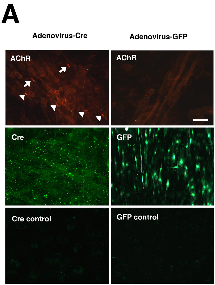
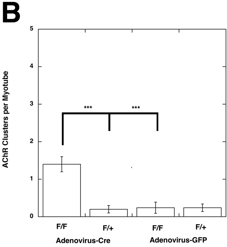
Figure 6. Induction of AChR clustering in primary myotubes after deletion of Pofut1 in vivo.
(A) Primary myotube cultures were made from mice homozyogous for a floxed allele of Pofut1 (F/F). Myotube cultures were incubated with saturating titers of Adenovirus bearing a cDNA for Cre recombinase (Adenovirus-Cre) or Adenovirus bearing a cDNA for GFP (Adenovirus-GFP). Adenovirus–Cre led to uniform, abundant, Cre expression in the majority of myotubes (middle left panel, with control (no primary antibody) lower left), and stimulated formation of both large (arrows) and small (arrowheads) acetylcholine receptor (AChR) aggregates, as evidenced by rhodamine-α-bungarotoxin staining (upper left). Adenovirus-Cre also increased overall bungarotoxin staining of myotube membranes. Adenovirus-GFP led to similar levels of infectivity (middle right), but did not increase AChR aggregates (upper right). Bar is 100µm. (B) Myotubes homozygous for floxed Pofut1 (F/F) that were infected with Adenovirus-Cre had significantly elevated levels of AChR clusters compared to similarly infected myotubes that were hemizyogous for floxed Pofut1 (F/+) or to F/F or F/+ cultures infected with Adenovirus-GFP. Errors are SEM for n=5–6 experiments.


