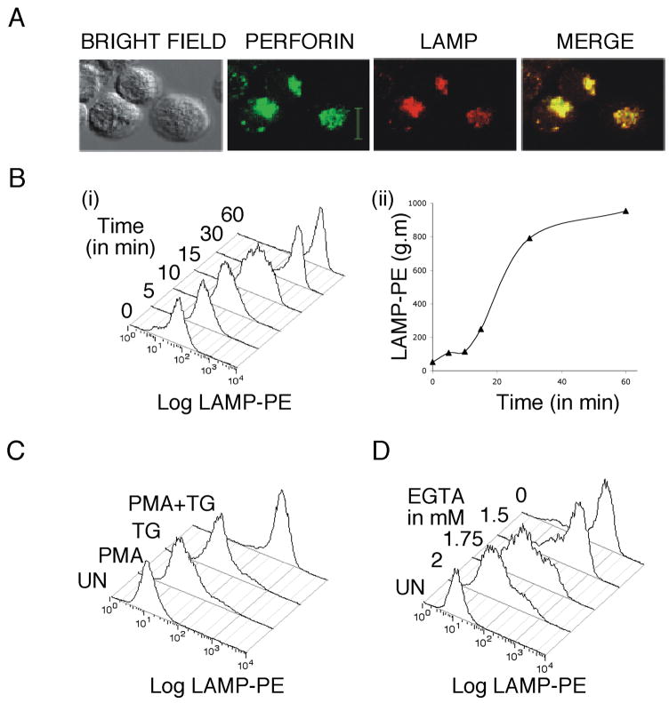
Figure 1. LAMP externalization as a marker for NK-92 lytic granule exocytosis.
A) Unstimulated NK-92 cells were fixed and permeabilized and stained for LAMP-1 and perforin. Scale bar denotes 10 μM. B) i) NK-92 cells were stimulated with TG and PMA and LAMP externalization was measured at various time points. Histograms of anti-LAMP fluorescence are shown. ii) Geometric mean (g.m.) from the experiment in (i) plotted vs. time. C) Representative histograms of anti-LAMP staining intensity showing the effects of stimulating cells with TG, PMA or both TG+PMA on granule release. D) Representative histograms of anti-LAMP staining intensity showing the effects of varying calcium levels on lytic granule exocytosis.

