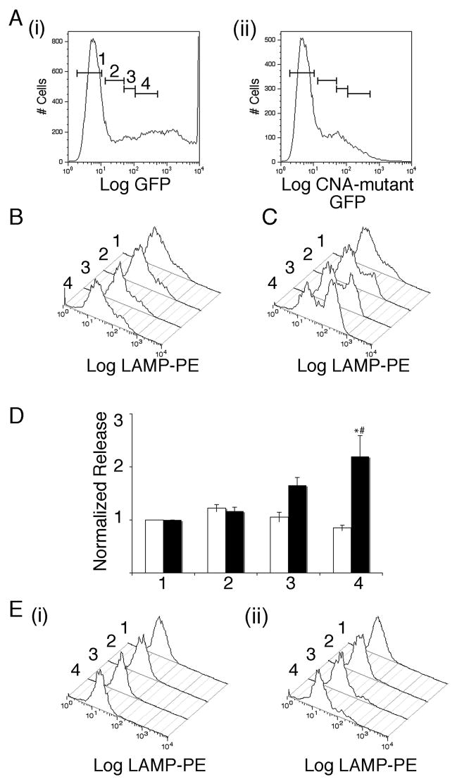
Figure 3. Effects of expressing mutant constitutively active calcium-independent calcineurin A on NK-92 lytic granule exocytosis.
A i) Representative plots of GFP fluorescence for cells transfected with GFP. The bars indicate gating regions used in analyzing LAMP externalization in panels below. ii) Representative plots of mutant calcineurin construct fused to GFP. B) Representative histograms of LAMP fluorescence for cells transfected with GFP and then stimulated with TG and PMA in medium supplemented with 1.75 mM EGTA for the various gating regions shown in A. Histograms are arranged with untransfected cells at the back and moving to front corresponds to increasing levels of expression. C) As in B for cells transfected with mutant calcineurin. D) Normalized release for four such experiments for the cells in the different gating regions. White bars denote GFP transfected cells and black bars denote mutant calcineurin transfected cells. Data were normalized to the level of release observed in stimulated cells from the GFP negative population. Normalized LAMP fluorescence values of mutant-calcineurin transfected cells that differ at p<0.05 level from untransfected cells is indicated by * and those that differ from equally GFP expressing cells is indicated by #. E) (i) Representative histograms of LAMP fluorescence for cells transfected with GFP and then stimulated with TG and PMA in 10mM EGTA containing medium for the various gating regions.(ii) As in (i) but for cells transfected with mutant calcineurin.

