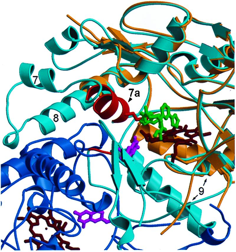
Figure 6.

Comparison of the 2–iNOS Δ114 complex with the murine iNOS oxygenase domain dimer. Compound 2 occupies the arginine-binding site. The monomers of the iNOS dimer structure (11) are shown in dark and light blue. The 2–iNOS Δ 114 structure (tan) has been aligned with a monomer (light blue) of the dimer structure; the hemes are shown in dark red. The iNOS Δ 114 heme and the dimer heme align very closely (for clarity only the iNOS Δ 114 heme is shown). Compound 2 (dark gray) displaces helix 7a (red) side chains from the arginine-binding site in the 2–iNOS Δ 114 structure, e.g., Glu-371 (red). The region from the beginning of helix 7a to the middle half of helix 8 is disordered in 2–iNOS Δ 114 structure.
