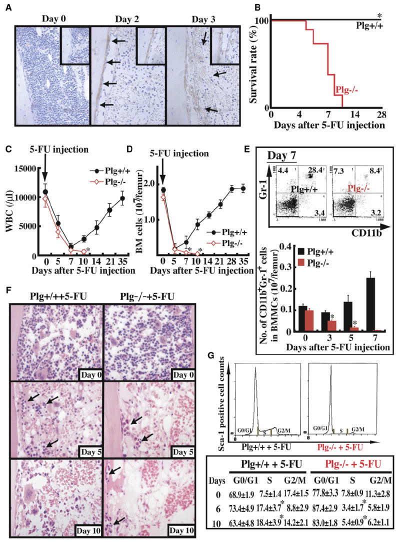
Figure 1. Delayed Bone Marrow Cell Recovery in Plg−/− Mice after Myelosuppression.

(A–G) Plg+/+ (n = 12) and Plg−/− mice (n = 12) received a single dose of the myelosuppressive agent 5-FU i.v.
(A) Immunohistochemical staining for Plg/plasmin (brown staining) in bone marrow (BM) sections from Plg+/+ and Plg−/− mice treated with 5-FU. Magnification ×100.
(B) Survival of 5-FU-treated mice was assessed daily.
(C) WBC counts were counted. Error bars represent standard error of the mean (SEM).
(D) Total number of BMMCs per femur was assessed. Error bars represent SEM.
(E) BM cells were stained for the myeloid markers CD11b-FITC and Gr-1-PE and analyzed by FACS (upper panel). Absolute numbers of CD11b+/Gr-1+ BM cells per femur were calculated (lower panel). Error bars represent SEM.
(F) H&E staining of BM sections after 5-FU treatment. Arrows depict hematopoietic cells. Magnification ×200.
(G) DNA content of Sca-1+ BM cells was determined after 5-FU treatment (n = 3 per group and time point). *p < 0.05.
