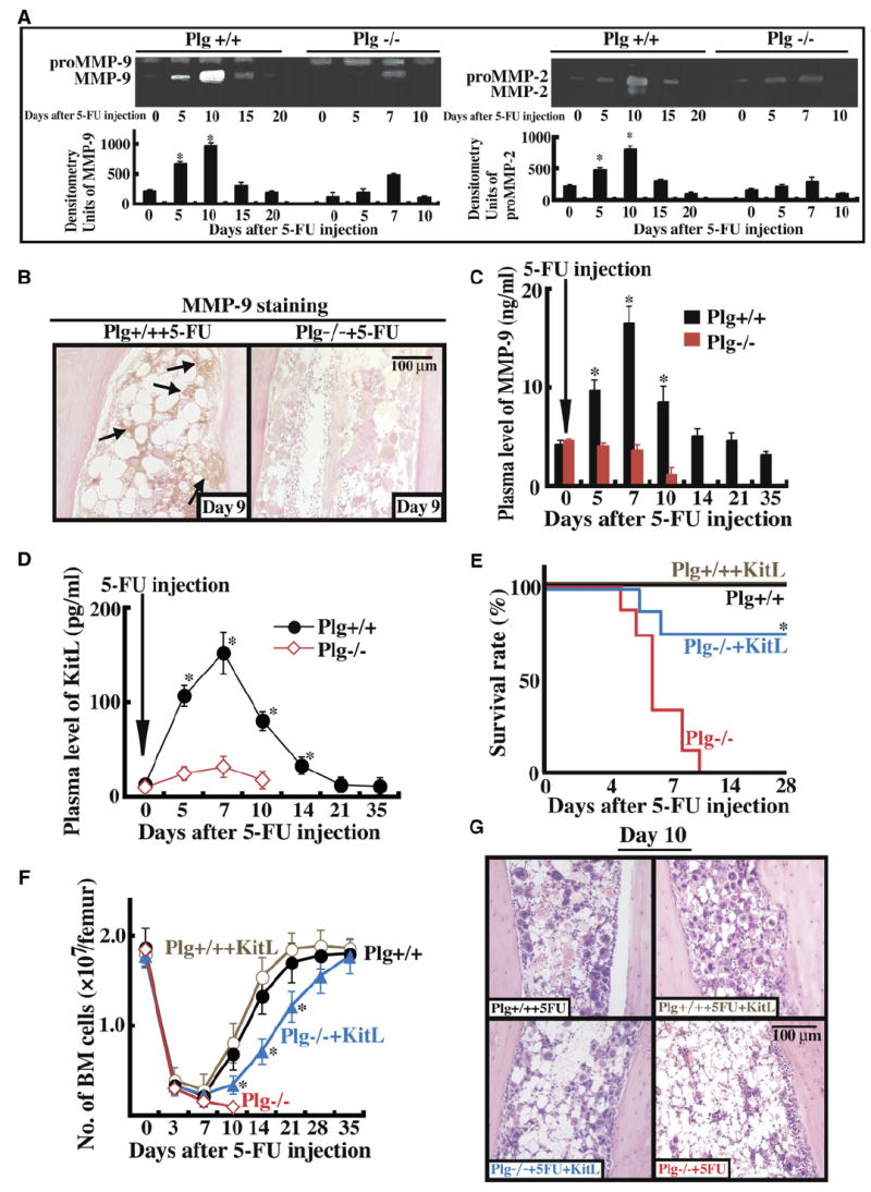
Figure 2. MMP Activation and KitL Release Are Impaired in Plg−/− Mice after Myelosuppression, Resulting in BM Recovery Failure.

(A–D) Plg+/+ and Plg−/− mice were injected with a single dose of 5-FU i.v. (A) BM cells (three mice per time point) were cultured in serum-free medium overnight. Cell supernatants were assayed for proMMP-9 (103 kDa), active MMP-9 (86 kDa), proMMP-2 (72 kDa), and active MMP-2 (62 kDa) by gelatin zymography. Error bars represent standard deviation. (B) Immunohistochemistry of BM sections 9 days after 5-FU injection for proMMP-9 with positive staining in the BM stromal compartment of Plg+/+, but less in the BM stromal compartment of Plg−/− mice. Magnification ×200. (C and D) Plasma obtained from peripheral blood (PB) was assayed for active MMP-9 (C) or KitL (D) by ELISA (p < 0.05). Error bars represent SEM. (E–G) Plg+/+ and Plg−/− mice were left untreated (n = 8) or received a single injection of 5-FU i.v. with/without daily i.v. injections of recombinant KitL (n = 6). (E) Survival of treated animals was determined daily. (F) Total number of BM cells per femur was assessed. Error bars represent SEM. (G) H&E staining of BM sections 10 days after 5-FU treatment. Magnification ×200. *p < 0.05.
