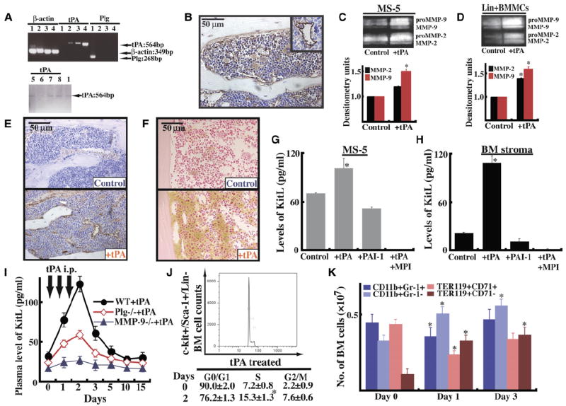
Figure 3. tPA-Mediated MMP Activation Releases KitL from Stromal Cells.

(A) RT-PCR for tPA and Plg in liver (sample 1; positive control for Plg), 4-week-old BM stroma of Plg+/+ mice (sample 2) and of Plg−/− mice (sample 3), MS-5 cells (sample 4), freshly isolated BM-derived lin− cells from Plg+/+ mice (sample 5), or Plg−/− mice (sample 6), as well as freshly isolated BM-derived lin+ cells from Plg+/+ mice (sample 7) or Plg−/− mice (sample 8). Agarose gel of one representative experiment.
(B) Immunohistochemistry for tPA in BM sections of Plg+/+ mice under steady-state conditions (magnification ×200). (Insert) Vessel stained positive for tPA.
(C–F) MS-5 cells (C) or lin+ BMMCs from Plg+/+ mice (D) were cultured overnight with/without tPA under serum-free conditions. Supernatants were analyzed by zymography for MMP-2 and MMP-9. Error bars represent standard deviation. Immunohistochemistry for Plg (E) and MMP-9 (F) in BM sections of Plg+/+ mice 3 days after starting tPA treatment (magnification ×200).
(G and H) Confluent MS-5 stromal cells (G) or Plg+/+ primary BM stroma cells (H) were cultured overnight in serum-free medium (n = 3) in the presence of recombinant tPA, recombinant PAI-1, and MPI (CGS 27023A) with or without tPA. Supernatants were collected and analyzed for KitL by ELISA (n = 3, p < 0.05). Error bars represent SEM.
(I–K) Plg−/− and MMP-9−/− mice and littermate mice were injected with a single dose of tPA or PBS daily from day 0–2 i.p. (I) Blood was drawn after tPA administration as indicated. Plasma samples were analyzed for KitL by ELISA (n = 6; ELISA was performed twice). Error bars represent SEM. (J) BM cells were collected on day 0 and day 2. Sca-1/ c-Kit+/Lin− (KSL) cells were costained with PI. Cell-cycle analysis was performed on KSL cells (n = 3). (K) BMMCs of tPA- and non-tPA-treated animals (n = 3) were incubated with anti-Ter119 and anti-CD71 antibodies. Proerythroblasts stained positive for CD71hiTer119hi, whereas erythroblasts stained positive for CD71loTer119hi. Granulocytic cells are positive for CD11b and Gr-1, whereas BM-derived monocytic cells showed positive staining for CD11b and expressed little or no Gr-1. Absolute numbers are given. Error bars represent SEM.
