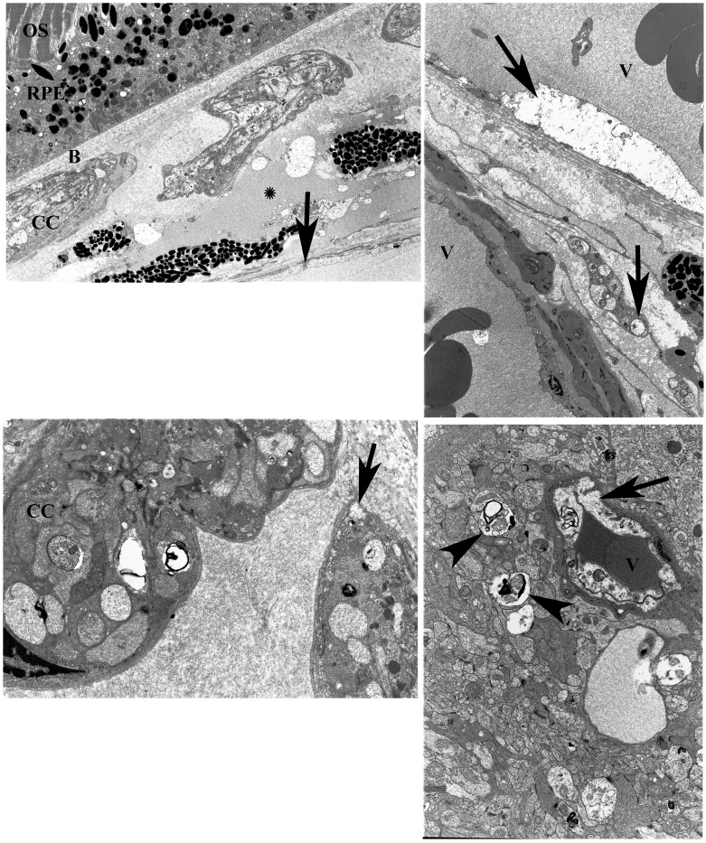
FIGURE 9.
Top left, In lesion K, the area of retina irradiated with 1800mW/cm2 with dye, outer segments (OS) appear normal, while the RPE (RPE) shows some vacuolation and loss of basal infoldings. The choriocapillaris (CC) is closed by platelet thrombus. The matrix shows plasma infiltration (asterisk) and damaged cells with debris. A larger vessel at the bottom shows endothelial disruption (arrow). No evidence of collagen denaturation. (uranyl acetate and Sato’s lead stain, original magnification ×2200). Bruch’s membrane (B). Top right, In lesion K, deeper choroidal vessels (V) show damage to the endothelium (arrow) and vacuolation to matrix cells (arrow), but no denatured collagen is seen (uranyl acetate and Sato’s lead stain, original magnification ×2950). Lower left, Higher magnification of lesion K shows closure of two choriocapillaries (CC) by platelets and fibrin with endothelial damage (arrow). The matrix shows normal collagen. (uranyl acetate and Sato’s lead stain, original magnification ×6610). Lower right, Retinal vessels are affected at 1800 mW/cm2 irradiance. This capillary (V) in lesion K shows damaged endothelium (arrow) and some of the surrounding nerve fibers are also affected (arrowheads) (uranyl acetate and Sato’s lead stain, original magnification ×5200).

