FIGURE 1.
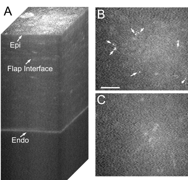
Confocal images taken after IntraLASIK from a quiet eye. A, Reconstruction of confocal microscopy through focusing (CMTF) stack. Only a slight increase in reflectivity is detected at the interface. B, Single image taken from the CMTF stack in A, at the flap interface. No keratocyte activation is observed, and interface particles are present (arrows). Bar indicates 75 μm. C, Single image taken 20 μm above the interface showing normal quiescent keratocytes. Epi, epithelial surface; Endo, endothelium.

