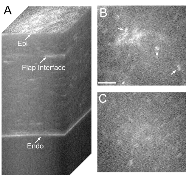
FIGURE 2.
Confocal images taken after IntraLASIK from an eye with keratocyte activation. A, Reconstruction of confocal microscopy through focusing (CMTF) stack. Note the areas of increased reflectivity at the flap interface. B, Single image taken from the CMTF stack in A, at the level of the flap interface. Note highly reflective nuclei (arrows), indicating keratocyte activation. Bar indicates 75 μm. C, Single image from the CMTF stack taken 20 μm above the flap interface. Quiescent keratocytes are observed. Epi, epithelial surface; Endo, endothelium.

