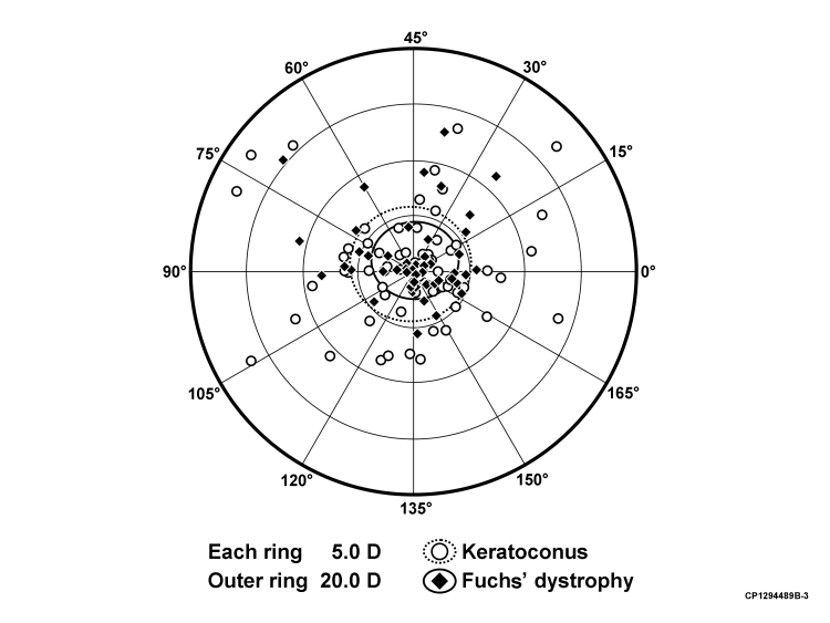
FIGURE 5.
Doubled-angle plot of the vectoral change (difference vector) observed in the astigmatism magnitude and axis for each subject between 13 months and last follow-up (if at least 15 years) after penetrating keratoplasty for keratoconus and Fuchs' dystrophy. The refractive centroid was 0.9 D × 51 ± 5.5 D, and the shape factor was ρ = 0.84 for keratoconus patients. The refractive centroid was 1.0 D × 44 ± 3.5 D, and the shape factor was ρ = 0.83 for Fuchs dystrophy patients. This suggests no difference in astigmatic shift in any given axis direction when comparing PK for keratoconus with PK for Fuchs' dystrophy.

