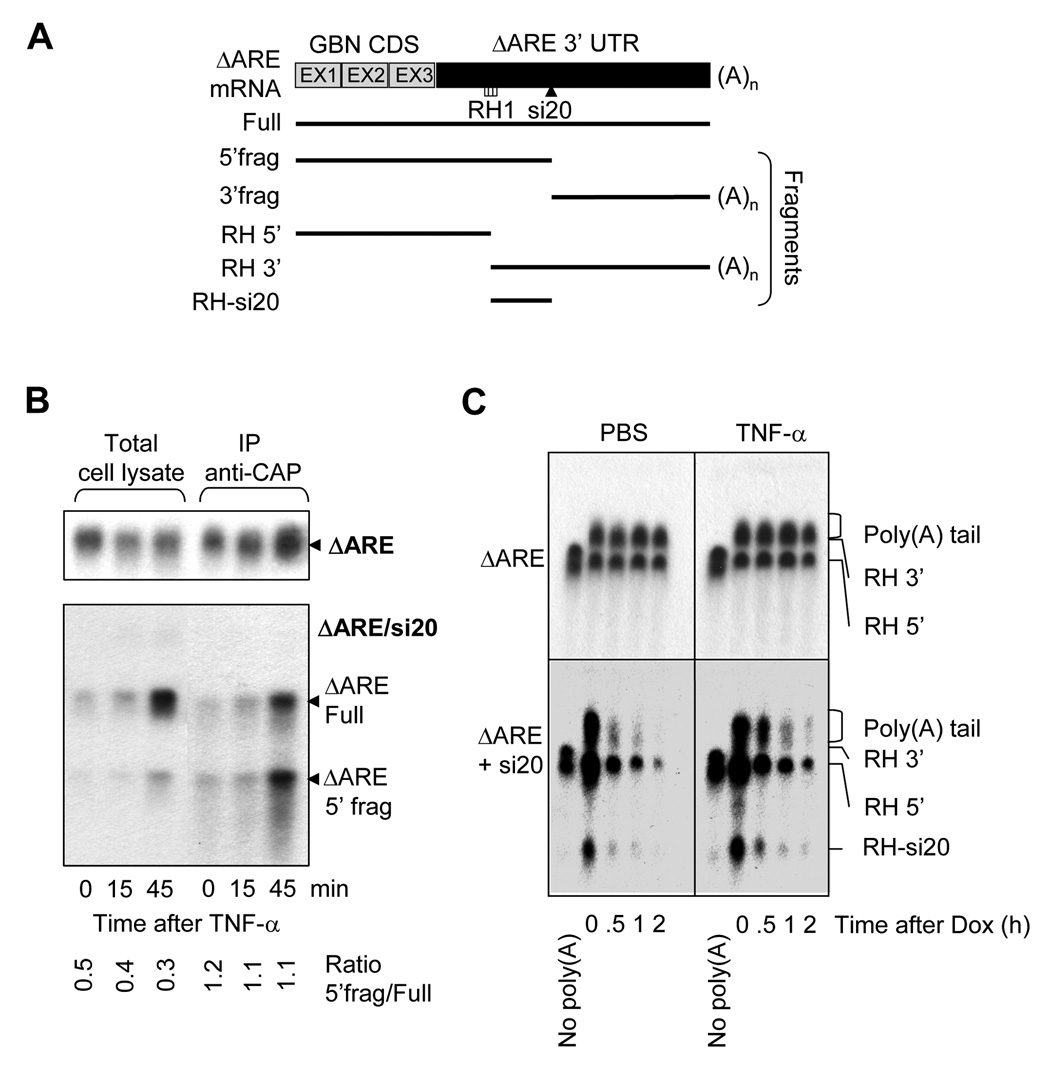
Figure 5. Capping and Poly(A) tail of si20 targeted ΔARE mRNAs after TNF-α stimulation.
A)Schematic representation of the fragments generated by RNAse H during ΔARE mRNA poly(A) tail analysis. B) HEK-293T cells were transfected with CMV-31 ΔARE (ΔARE), or CMV-ΔARE plus pSuper-si20 (ΔARE/si20). Cells were treated with TNF-α for 0, 15, or 45 min. and total RNA was isolated. Cap-containing mRNAs were immunoprecipitated with K121 anti-2,2,7-trimethylguanosine agarose-conjugated mouse monoclonal antibodies. Northern-blotting was done with RNA extracted from cell lysates and after K121 immunoprecipitation. C) HeLa-Tet-off cells were transfected with Tet-ΔARE (ΔARE), or Tet-ΔARE plus pSuper-si20 (ΔARE/si20). Cells were treated for 15 min. with PBS or TNF-α prior to doxycycline addition for 0, 0.5, 1, and 2 hours. RNA was extracted and subjected to RNAse H treatment to visualize poly(A) tail length. As a control of deadenylation, oligo d(T)20 was added to remove the poly(A) tail. Northern-blots were used to analyze poly (A) tail distributions.

