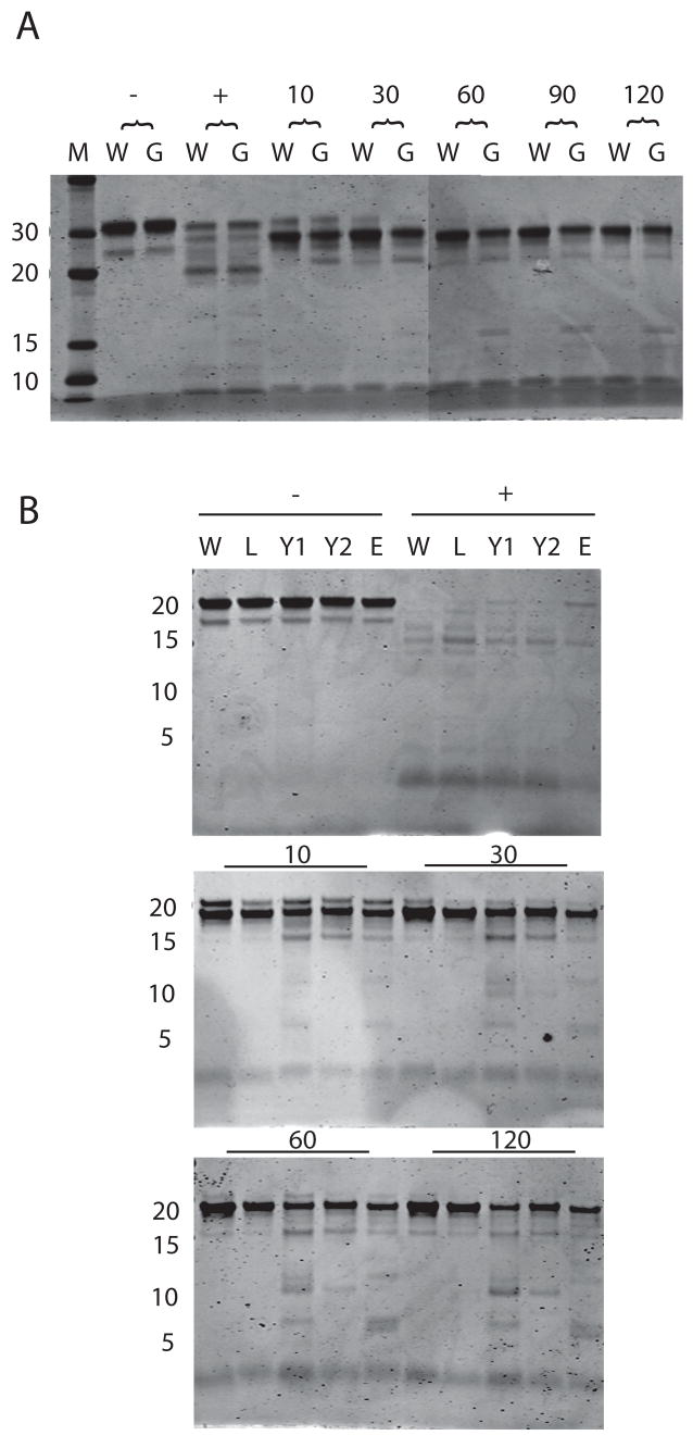
Figure 3. Protease sensitivity of WT PrfA and PrfA* mutants.
A. Wild type (W) and PrfA G145S (G) recombinant proteins were digested for 10, 30, 60, 90, and 120 minutes with trypsin, subjected to SDS-PAGE and visualized by Coomassie stain. Minus symbol denotes protein without trypsin, plus symbol denotes trypsin digestion of denatured protein. Lane M contains molecular weight markers. B. Wild type and PrfA* proteins were treated with trypsin for 10, 30, 60, and 120 minutes. W, wild type; L, L140F; Y1, Y63C, Y2, Y154C; E, E77K. Minus symbol indicates samples without trypsin, plus symbol indicates trypsin treated denatured protein. Numbers on left represent molecular weight in kD. Gel is representative of three similar experiments.

