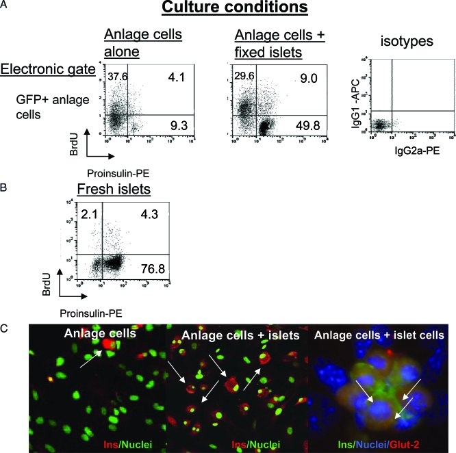
Figure 1.
Induction of proinsulin in anlage cells cultured with fixed adult islets: A, Proinsulin expression and BrdU labeling was analyzed by flow cytometry. Cells from 7 d cultures of pancreatic anlage from GFP+ mice were intracellularly stained with fluorescently labeled mAbs to proinsulin and BrdU. Dot plots of the FACS staining are shown. Gating was placed on GFP+ anlage cells. The percentage of cells in quadrants are shown. The staining with control isotype antibodies is shown at the far right. B, Comparative staining of freshly isolated islets. C, Representative immunohistochemical staining of insulin in cells from cultures of anlage alone (left) or anlage with adult islets (middle). Proinsulin+GFP+ cells are identified with arrows. The panel on the far right is from another experiment in which anlage cells from nontransgenic mice were cocultured with islet cells and stained for insulin and Glut-2 (arrows). The cells from the mature islets that had been permeabilized, fixed, and stored did not stain with antibody to insulin. PE, Phycoerythrin.

