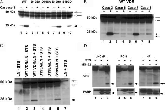
Figure 3.
Cleavage of the VDR at the caspase-3 site in vitro and in intact cells. A, COS-7 cell extracts expressing the WT VDR and mutant VDRs were incubated with 1 U caspase-3 for 1 h at 37 C. Cleavage products were then detected by immunoblot using anti-VDR D-6 antibody. B, Differential sensitivity of VDR to caspases in vitro. Recombinant human caspase-3, -6, -7, and -8 (1 U each) were incubated with COS-7-expressed WT VDR for 1 h at 37 C and then analyzed by immunoblot with anti-VDR D-6 antibody. C, Extracts from naive and STS-treated LNCaP cells were incubated with COS-7 cell extracts expressing the WT VDR and mutant VDRs for 1 h at 37 C. Cleavage products were then detected by immunoblot using anti-VDR D-6 antibody. LN, LNCaP cell extracts. D, LNCaP and PC-3 prostate cancer cells and human skin fibroblasts were treated with 1 μm STS for 3 h in the absence and presence of 10 μm MG-132. VDR cleavage products were then detected by immunoblot using anti-VDR D-6 and PARP cleavage detected using anti-PARP antibodies. Gray arrows show intact VDR. Black arrows show the VDR cleavage fragment. White arrows indicate nonspecific bands. Arrowhead indicates PARP cleaved fragment. HF, Human fibroblasts.

