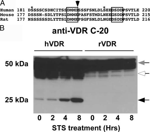
Figure 4.
The aspartic acid at the critical P1 position in the human VDR is not conserved in the mouse and rat VDRs. A, Alignment of human, mouse, and rat VDR sequences surrounding caspase-3 site. Asterisks indicate phosphorylation sites (S182 and S208), arrow indicates caspase-3 cleavage site, and boxes indicate DxxD motifs. B, COS-7 cells expressing the human or rat WT VDRs were treated with 1 μm STS and samples taken at the indicated times. Protein extracts were subjected to SDS-PAGE and immunoblotted with a VDR antibody (anti-VDR C-20) that recognizes the C terminus of human and rat VDRs. Intact VDR migrates as approximately 50 kDa (gray arrow). Black arrows indicate the VDR cleavage fragments. White arrows indicate nonspecific bands.

