Figure 3.
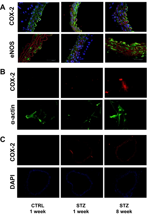
Expression of COX-2 and eNOS is increased in aortas from mice with STZ-induced diabetes. Aortas from mice treated with CTRL or STZ for the indicated times were prepared as described in Materials and Methods. A, Aortas were fixed in 8% paraformaldehyde and ring sections (10 μm) were immunolabeled with antibodies against COX-2 (upper panel) or eNOS (lower panel). Sections were immunostained with IgG-fluorescein isothiocyanate conjugated- (upper panel) and IgG-Texas Red conjugated (lower panel)-laminin antibodies. Finally, DAPI staining was used to visualize cell nuclei. Thus, visualization by confocal microscopy shows red fluorescence for COX-2 staining and green fluorescence for laminin staining in the upper panel. In the lower panel, green fluorescence is seen for eNOS staining, whereas red fluorescence is seen for laminin staining. Cell nuclei are visualized in blue. Representative results are shown for experiments independently repeated three times. B, VSMCs were prepared from aortas of mice treated with CTRL or STZ for the indicated times as described in Materials and Methods. VSMCs were fixed in 3.5% paraformaldehyde and immunolabeled with antibodies against COX-2 (upper panel) or α-actin (lower panel). Representative results are shown for experiments that were repeated independently three times. C, Mesenteric arteries were fixed in 8% paraformaldehyde and ring sections (8 μm) were immunolabeled with COX-2 antibody (upper panel). DAPI staining was used to visualize cell nuclei (lower panel). Red fluorescence indicates COX-2 staining. Cell nuclei are visualized in blue. Representative results are shown for experiments independently repeated three times.

