Figure 2.
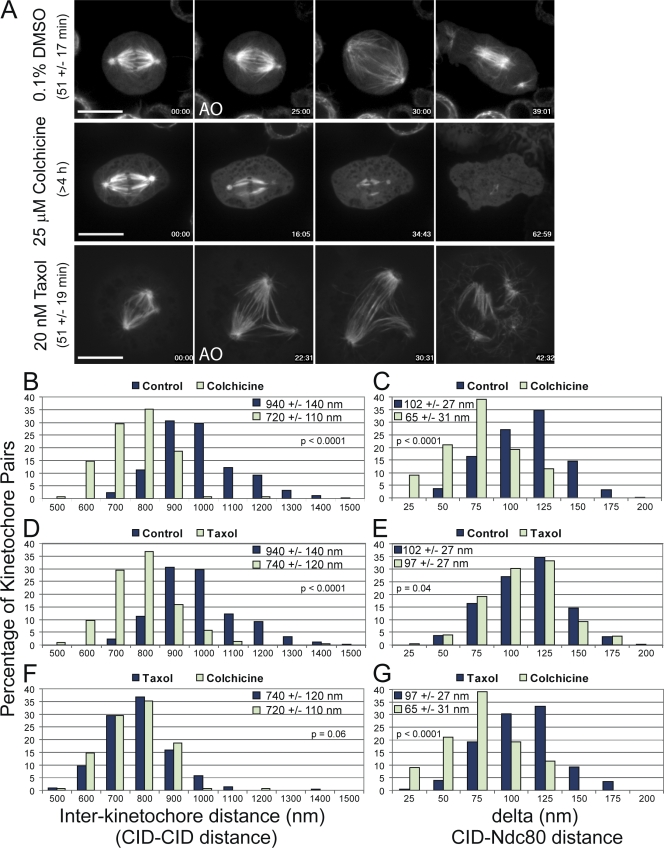
Microtubule poisons differentially affect interkinetochore distance and delta. (A) Selected frames from time-lapse imaging of GFP-tubulin–expressing S2 cells after treatment with DMSO, 25 µM colchicine, or 20 nM taxol. DMSO control cells progress through mitosis (as defined by chromosome condensation to anaphase onset [AO]) in 51 ± 17 min (n = 124 cells). Spindle microtubules completely depolymerize within 1 h after the addition of 25 µM colchicine, which causes cells to delay in mitosis for at least 4 h (n = 40 cells). Cells treated with 20 nM taxol progress through mitosis with the same kinetics as control cells (51 ± 19 min; n = 121 cells). (B–G) The distributions of interkinetochore distances (left) and delta (right) values are shown for all treatments. (B and C) Colchicine treatment causes reduction of both interkinetochore distance and delta, defining the rest lengths for each parameter (n = 156 kinetochore pairs). (D and E) Taxol treatment (n = 228 kinetochore pairs) causes a reduction in the interkinetochore distance; however, delta is not dramatically reduced relative to controls (n = 346 kinetochore pairs). (F and G) Comparing taxol and colchicine treatments highlights the fact that 20 nM taxol causes the interkinetochore distance to approach rest length without dramatically reducing delta. The mean values ± standard deviations and the two-tailed p-values for all conditions are shown in each graph. Bars, 10 µm.

