Figure 1.
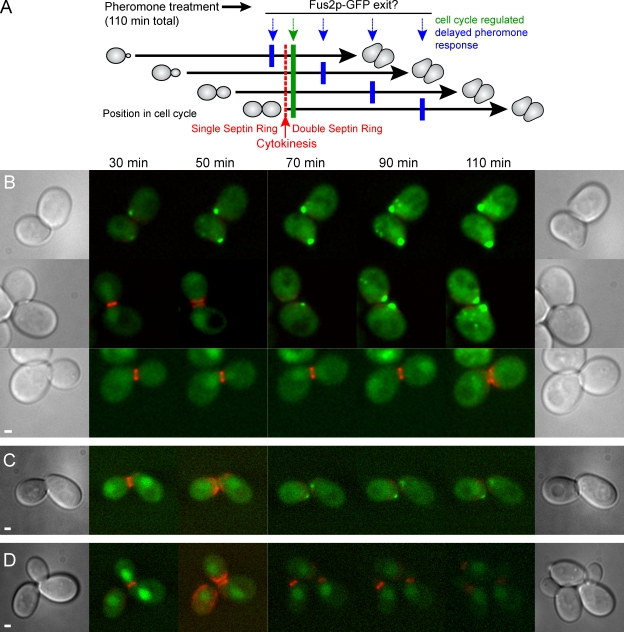
Fus2p is expressed in pheromone-treated cells before cytokinesis but remains in the nucleus until after cell division. (A) Experiment schematic. An asynchronous cell population expressing Fus2p-GFP and Cdc3-mCherryFP, an indicator of cytokinesis (red), was treated with pheromone for 110 min (black arrows) and examined microscopically over time. If Fus2p-GFP nuclear exit is solely dependent on pheromone signaling, exit should occur at similar times after exposure for all cells (blue arrows). If Fus2p-GFP nuclear exit requires completion of the previous cell cycle, exit should occur soon after cytokinesis independent of the length of pheromone signaling (green arrow). (B) Fus2p-GFP expressed under its own promoter. Strain MY10176 containing FUS2∷GFP104 and CDC3-mCherry was pregrown in selective medium, and α factor was added at t = 0. Representative cells are shown in which cytokinesis occurred before 30 min, between 30 and 50 min, and between 90 and 110 min (top, middle, and bottom, respectively). (C) Fus2p-GFP expressed under the GAL1 promoter. Strain MY10177 containing PGAL1-FUS2∷GFP104 and CDC3-mCherry was pregrown in selective medium with galactose; α factor and glucose were added at t = 0. (D) Pheromone is required for Fus2p-GFP exit G1. MY10177 was examined as in C except that no α factor was added. Bars, 1 µm.

