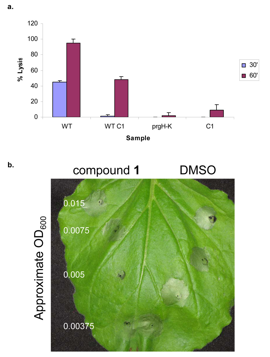
Figure 5. Inhibition of bacterial virulence phenotypes by compound 1.
a, Mouse bone marrow macrophages (BMM) from Balb/c mice were infected with WT S. typhimurium grown in the absence (WT) or presence of 380 µM of compound 1 (WT C1) to SPI1 inducing conditions. A T3S genetic mutant (prgH–K) was used as a negative control. To confirm that compound 1 was not itself cytotoxic to the macrophages, compound without bacteria (C1) was added at a concentration equal to the experimental samples. Bacteria were added at an MOI of 40 and cytotoxicity was assessed by LDH release at 30 minutes and 60 minutes after infection. Assays were performed in quadruplicate and mean standard deviations are shown. b, P. syringae and compound 1 were co-inoculated on non-host tobacco plants and monitored for HR. Varying concentrations of bacteria were added, while the amount of compound or solvent (v/v) remained constant. Four independent experiments were performed and a representative experiment is shown. Tissue collapse near the point of inoculation for either compound 1 or solvent alone is due to tissue damage caused during injection of compounds by a blunt-ended syringe.

