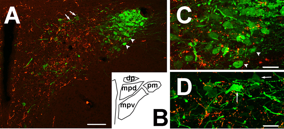
Fig. 3. AVP-immunoreactive neurons and GLP-1-immunoreactive nerve fibers in the paraventricular nucleus.
A: AVP expressing cells are packed in the core of the posterior magnocellular division of the PVN. GLP-1 immunoreactivity (red) is mainly expressed in the medial parvocellular division (projection image). B: Schematic diagram of the approximate area shown in panel A. C: Higher magnification figure of the posterior magnocellular division of the PVN, illustrating sparse GLP-1 fiber distribution in the AVP-rich core of this area, along with only occasional instances of bouton-soma appositions (individual 0.5 um optical section). Arrowheads in A correspond to neurons noted in C. D: Higher magnification figure of the medial parvocellular division of the PVN (individual 0.5 um optical section). Note the scattered AVP positive neurons receiving GLP-1 innervation in this region. Arrows in A correspond to indicated cells in D. Scale bars: 100µm (A) and 30µm (C, D).

