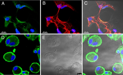
Fig. 5.
Tube-like structures formed by CD11b+ alveolar macrophages express LEC markers. After culturing in Matrigel for up to 31 days, cells were fixed, permeabilized, and stained by indirect immunofluorescence for LYVE-1 (green) (A) and podoplanin (red) (B). (C) A merged DIC and fluorescence image shows tube-like structures in the IPF samples that were reactive with LEC markers, anti–LYVE-1, and anti-podoplanin antibodies. (D–F) In contrast, cells from healthy volunteers show reactivity with anti–LYVE-1 antibodies outlining their periphery (D), without the formation of tube-like structures. Representative cells are shown in DIC and fluorescence-overlaid images. Cell nuclei were stained by DAPI (blue).

