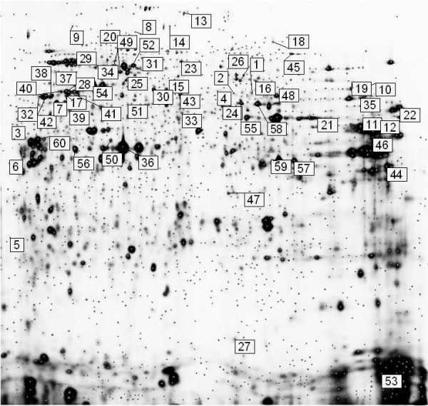
Figure 2.

Representative 2D DIGE gel image of Cy2-labelled pool of Clone #3 and Clone #8 cell lysate samples. Differentially expressed proteins that have been successfully identified by MALDI-TOF MS (p ≤ 0.05, protein fold ≥ 1.2) are represented on the gel using DeCyder software. Proteins are labelled numerically for visual clarity and are outlined in Table S1.
