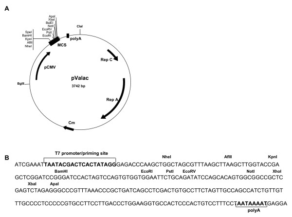
Figure 1.
Structure of pValac plasmid. A: Boxes indicate: Multiple cloning site (MCS) and BGH polyadenylation region (polyA). Arrows indicate: cytomegalovirus promoter (pCMV); replication origin of L. lactis (Rep A) and E. coli (Rep C) and chloramphenicol resistance gene (Cm). ClaI and BglII restriction sites used to ligate eukaryotic and prokaryotic regions are showed. B: Multiple cloning site showing the T7 promoter/priming site, different restriction enzyme sites and polyA site.

