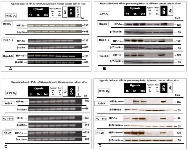
Figure 2.

HIF-1α-mRNA and -protein expression under extreme hypoxic (0.1% O2) and normoxia and reoxygenation conditions in HepG2, Hep 1-6, Hep3B, A-549, HT-29 and HCT-116 cells, in vitro. (A) Semi quantitative RT-PCR analysis of HIF-1α mRNA expression in HepG2, Hep 1-6 and Hep3B (B) Western blot analysis of HIF-1α protein expression under identical conditions in HepG2, Hep 1-6 and Hep3B. (C) Semi quantitative RT-PCR analysis of HIF-1α mRNA expression in A-549, HT-29 and HCT-116, in vitro (D) Western blot analysis of HIF-1α protein under identical conditions in A-549, HT-29 and HCT-116. Representative experiments out of three for each experimental set.
