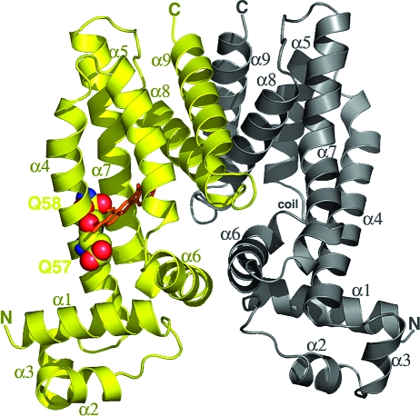
Figure 1.
Structure of the QacR(E58Q)−Be complex. A ribbon diagram of QacR(E58Q) bound to berberine with the drug-bound subunit colored yellow and the drug-free subunit gray. Secondary structural elements and the N- and C-termini are labeled. Residues E57 and E58 are shown as CPK. Berberine is shown as orange sticks.

