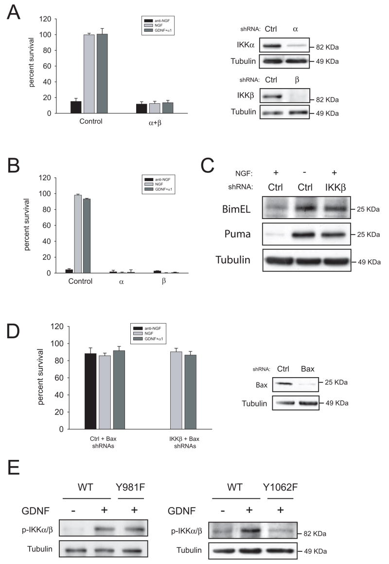
Figure 8.
IKKs are necessary for NGF- and GDNF-mediated survival of sympathetic neurons. (A and B) Neurons were infected with shRNAs to IKKs in combination (“α+ β”) or alone (“α” or “β”) two days after plating. After three additional days in the presence of NGF, cells were switched to the indicated factors and survival was evaluated 48 hours later. The right panel shows efficacy of the knockdown. (C) Silencing of IKKs induces BimEL and PUMA expression. Cells were infected the second day after plating with control shRNAs or a shRNA to IKKβ and kept on NGF for three additional days. Cells were then deprived or kept in NGF for 24 more hours as depicted, and BimEL and PUMA expression was assessed by western blot. (D) Silencing of Bax rescues cells from knockdown of IKKβ. Cells were infected with shRNAs to Bax, and subsequently infected with control or shRNAs to IKKβ as detailed in methods. Cells were maintained in NGF for three additional days, and switched to media containing the indicated factors. Cell survival was assessed 48 hours later. Right panels shows efficacy of the knockdown. (E) Loss of tyrosine 1062, but not tyrosine 981, blocks GDNF-mediated phosphorylation of IKKα/β. Sympathetic neurons form the indicated genotypes were cultured in the presence of NGF for five days, deprived overnight and stimulated for 10 minutes with GDNF. Blots were probed with antibodies to phospho-IKKs. Note that due to a higher acrylamide percentage IKKα and IKKβ were not resolved as in figure 7C.

