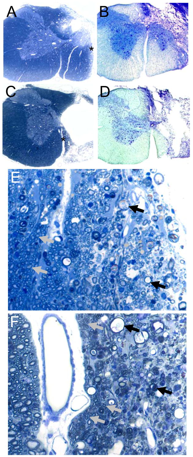
Figure 1. Representative histological examples of cervical hemilesions.
Panel A provides an example of moderate VM white matter sparing in a semi-thin (2 μm) plastic section stained with toluidine blue. Panel B shows an example of moderate VM sparing as assessed in a thicker (40 μm) cryostat section stained with luxol fast blue and cresyl violet. Panels C and D depict minimal VM sparing in a similarly stained semi-thin plastic (C) and 40 μm cryostat section (D). Magnification for panels A-D is 2.5x. Panel E depicts a higher magnification (40x) of the lateral edge of the lesion site as indicated by the asterisk in panel A. The tissue at the right of panel E shows numerous axonal profiles at different stages of Wallerian degeneration (examples indicated by black arrows). Towards the left of panel E (i.e. towards the spinal midline) many apparently healthy axons can be observed (a few are indicated by gray arrows). Panel F is a higher magnification (40x) of the site indicated by the asterisk in panel C. Thus, the injured side is to the right and the intact contralateral side is to the left of the ventromedian fissure (the white area just the left of center). The “spared tissue” at the far right of the figure represents extensively degenerated white matter although a few small axons are present. More medially, the tissue in Panel F consists of numerous axonal profiles at different stages of Wallerian degeneration (black arrows). Some axonal preservation is seen, however, especially along the medial subpial surface adjacent to the ventromedian fissuer as indicated by clusters of small diameter myelinated axons (gray arrows).

