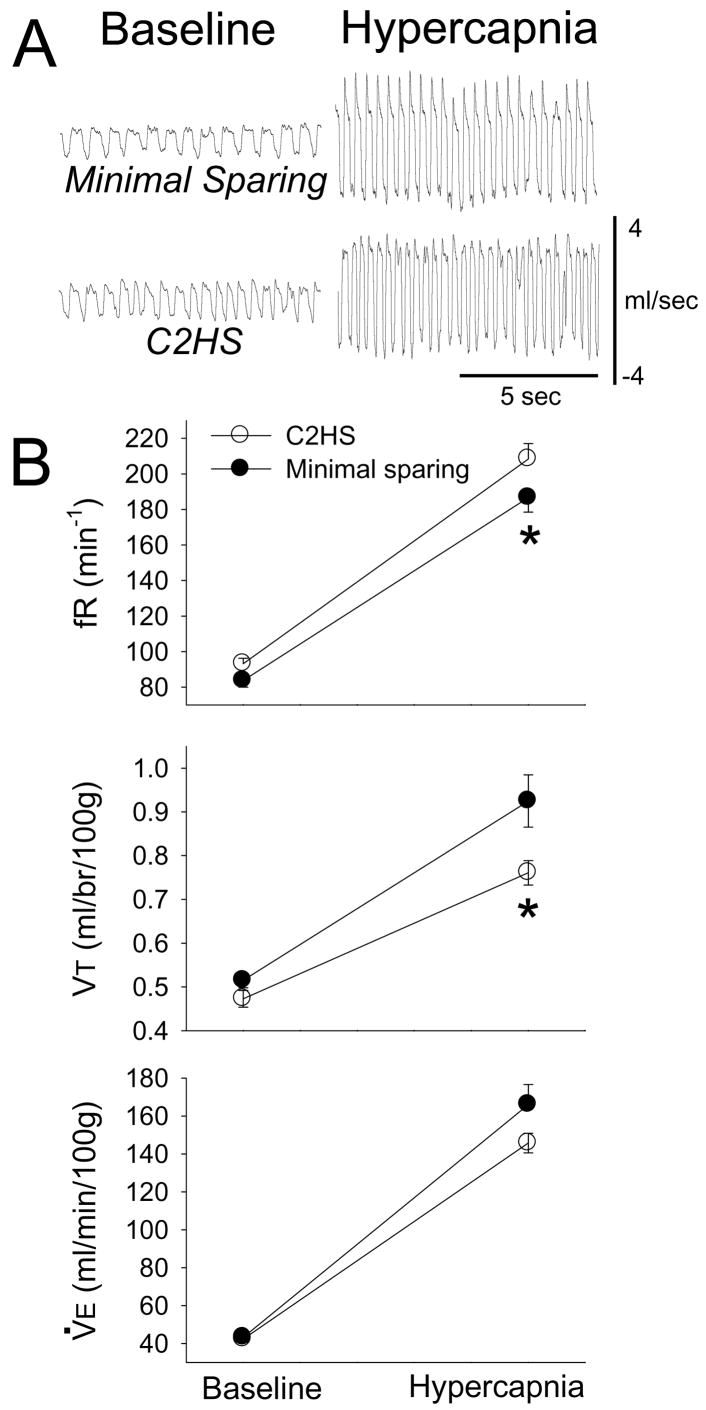
Figure 2. Ventilation during baseline and hypercapnic challenge (7% CO2) in rats with anatomically complete C2HS injury vs. minimal VM tissue sparing.
Panel A presents representative airflow traces depicting in breathing in unanesthetized rats. Scaling is identical in all panels. Panel B presents mean inspiratory frequency (fR), tidal volume (VT) and minute ventilation (V̇E). See text for description of 2-way repeated measures ANOVA results. *, significantly different than the C2HS group

