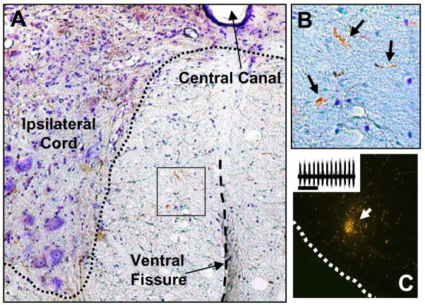
Figure 5. Example of anterogradely labeled projections in the VM medial cervical spinal cord.
Miniruby conjugated with biotin was delivered via iontophoresis into the brainstem (see methods). Panel A (10x magnification) shows the ventral horn of the cervical spinal cord ipsilateral to the lesion site. This rat had a “moderate VM sparing” lesion. Panel B provides a higher magnification (20x) view of the area indicated by the box in panel A. The arrows indicate processes that are BDA-positive (brown color). Panel C shows an example the medullary injection site. The inset in panel C shows the inspiratory bursting recorded at the injection site and just prior to iontophoresis.

