Figure 3.

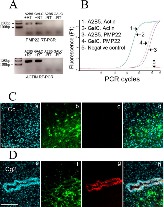
PMP22 is not translated in oligodendrocytes. A, PMP22 mRNA expression in oligodendrocytes. The RT-PCR shows the presence of PMP22 in A2B5+ GalC− cells and A2B5− GalC+ cells. The −RT lanes (without reverse transcriptase) are negative controls, and β-actin PCR is used as positive control. B, Quantification of PMP22 mRNA level. The PCR curves obtained with A2B5− GalC+ cDNA and A2B5+ GalC− cDNA show an increase of PMP22 mRNA level during differentiation. The overlapping curves for β-actin confirm the equivalent levels of mRNA in both samples. C, The CC1+ oligodendrocytes in the corpus callosum (Cc) are not immunoreactive for PMP22. Sagittal sections of postnatal rat brains are processed with a rabbit anti-PMP22 antibody (c) and the CC1 mouse antibody is used to label oligodendrocytes (b). Nuclei are visualized with DAPI (a). d, Merge picture. Scale bar, 100 μm. D, PMP22 protein is present in neuroepithelial cells. The CC1+ oligodendrocytes (f) in the cingulate cortex (Cg2 area) do not express PMP22 (g). A clear signal for PMP22 is obtained in neuroepithelial cells surrounding the ventricle. Nuclei are visualized with DAPI (e). h, Merge picture. Scale bar, 100 μm.
