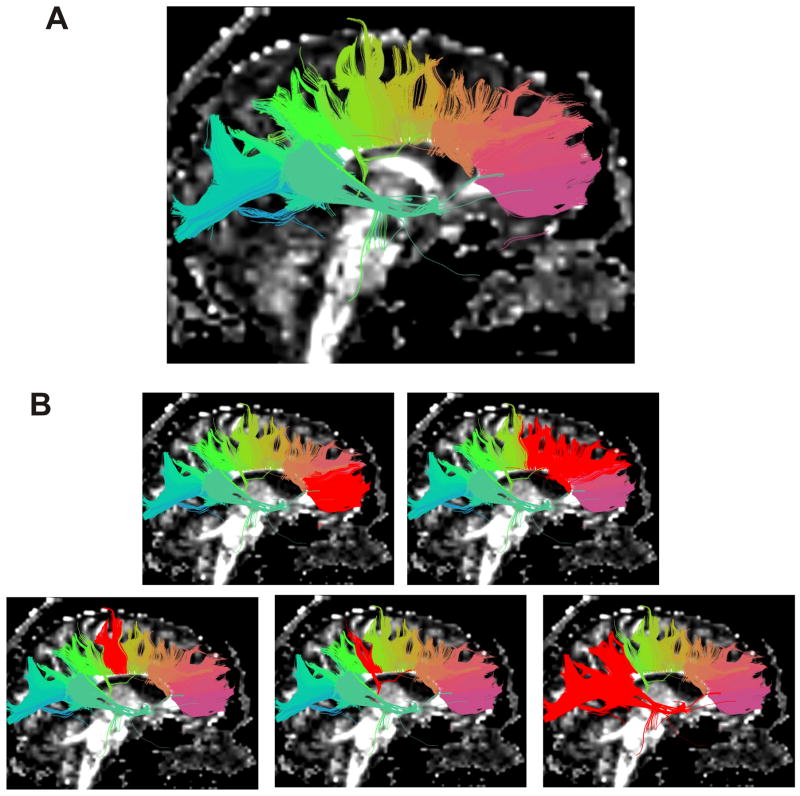
Figure 3.
(A,B). Segmentation of corpus callosum, and selection of subdivisions. Tracts are grouped into clusters according to similarity of shape and location and are colour coded accordingly (A). Clusters are then selected (B), red highlighted from left to right, as genu, premotor and supplementary motor projections, motor projections, sensory projections, and finally parietal temporal and occipital projections.

