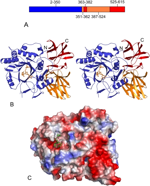
Figure 2. Structure of Endo-A.
(A) Diagrammatic representation of Endo-A. Amino acids 1–350 make up Domain 1 (blue), segments 351–362 and 387–524 make up Domain 2 (orange), while segments 363–386 and 525–611 make up Domain 3 (red). (B) Stereo image of Endo-A. Man3GlcNAc-thiazoline moiety is shown as sticks. (C) A surface electrostatic potential representation of Endo-A showing the Man3GlcNAc-thiazoline moiety sitting inside the active site cleft.

