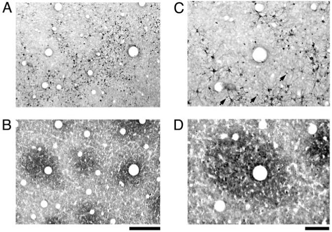
Figure 3.

High magnification photographs of SMI-32 immunoreactivity (A and C) and CO staining (B and D). The pattern of SMI-32 was comprised of dark neuronal somata and dendritic processes, as well as apical dendrites from deeper neurons that created a punctate pattern outside of blobs (arrows in B). At this magnification, individual SMI-32 positive cells could be seen to lie primarily outside of blobs. Scale bars = 250 μm (A and B) and 100 μm (C and D).
