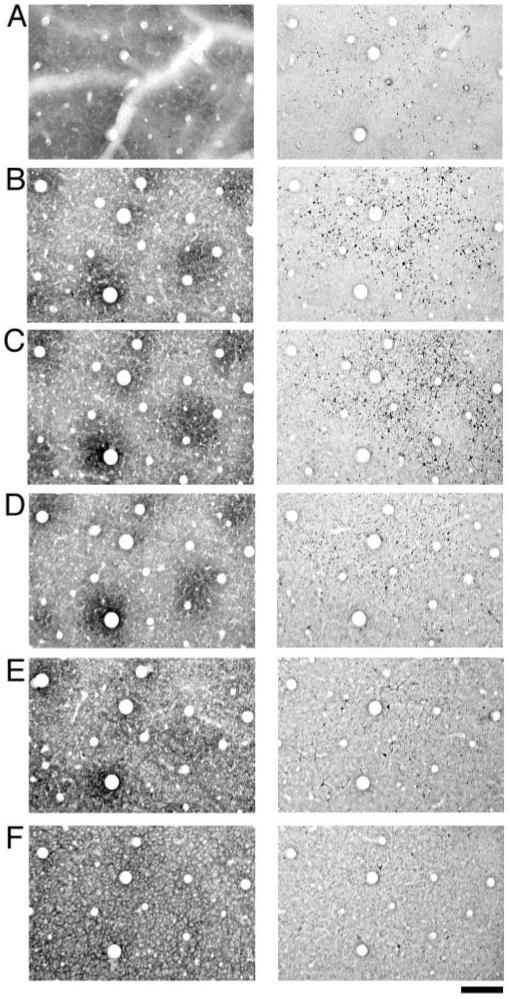
Figure 5.

A sequential series of sections reacted for CO (left column) or SMI-32 (right column). The sections start at the top of layer I (row A), move through layers II/III (rows B, C and D) and end in layer IVA (row F). SMI-32 labeling was apparent in the most superficial sections examined and consisted of apical dendrite shafts (row A). Sections taken from layers II/III (rows B, C and D) revealed a distribution of SMI-32 labeled cells that clustered outside of CO blobs. Sections sampled from deep layer III and layer IVA show weak SMI-32 labeling with the distribution of cells not showing a strong correlation with the CO pattern (E and F). Scale bar = 200 μm.
