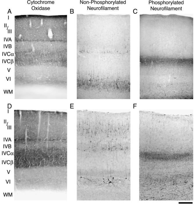
Figure 1.

Coronal sections of monkey V-1 reacted for CO, non-phosphorylated, and phosphorylated neurofilament from a normal 4 month old infant (A, B and C) and a normal adult (D, E and F). Non-phosphorylated neurofilament labeling in the infant (B) and adult (E) was strong in layers VI, V and IVB. Although there was heavy layer II/III reactivity in the adult, the infant showed light labeling. Phosphorylated neurofilament labeling was strong within layer IVCα in the infant (C) and adult (F) but comparatively weak in all other layers. Scale bar = 250 μm.
