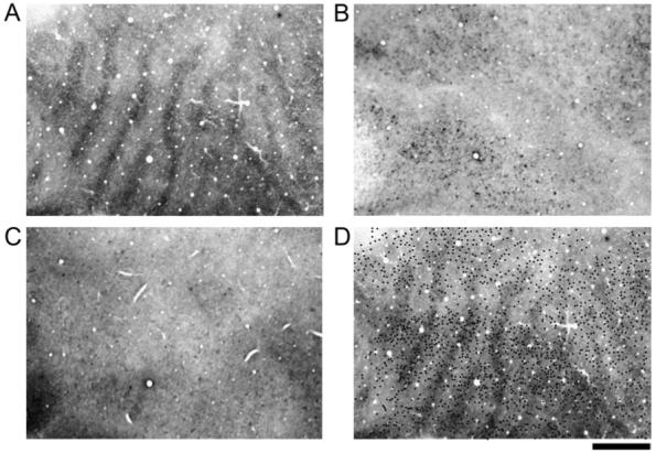
Figure 6.

The patterns of CO (A), non-phosphorylated neurofilament (B) and phosphorylated neurofilament (C) in V-1 of a monocularly deprived adult monkey (Mac 6). This monkey was deprived for 3 months at 7 years of age. The deprivation produced a low contrast pattern of dark and light CO bands throughout V-1 (A). Non-phosphorylated (B) and phosphorylated (C) neurofilament labeling was not banded after adult monocular deprivation. Non-phosphorylated labeled cell bodies were plotted and then superimposed onto the aligned pattern of CO (dots in D). Labeled neurons were roughly evenly distributed within both deprived and non-deprived regions of V-1. Adult monocular deprivation did not result in a loss of neurofilament labeling within V-1. Scale bar = 1 mm.
