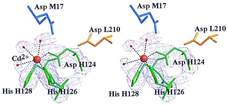
Figure 2.

Stereoview of the Cd2+ binding site (orange) on the RC from Rb. sphaeroides. The six Cd2+ ligands are His-H126, His-H128, Asp-H124 (green), and three water molecules (red). Two nearby aspartic acid residues, Asp-L210 (yellow) and Asp-M17 (blue), are part of a hydrogen bonding network that leads from the metal site to QB⨪ (see Fig. 5). |Fo| − |Fc| difference electron density (purple) is contoured at 2.5σ and superimposed on the structure. To reduce phase bias, ligands were excluded in the calculation of the map.
