
Fig. 1.
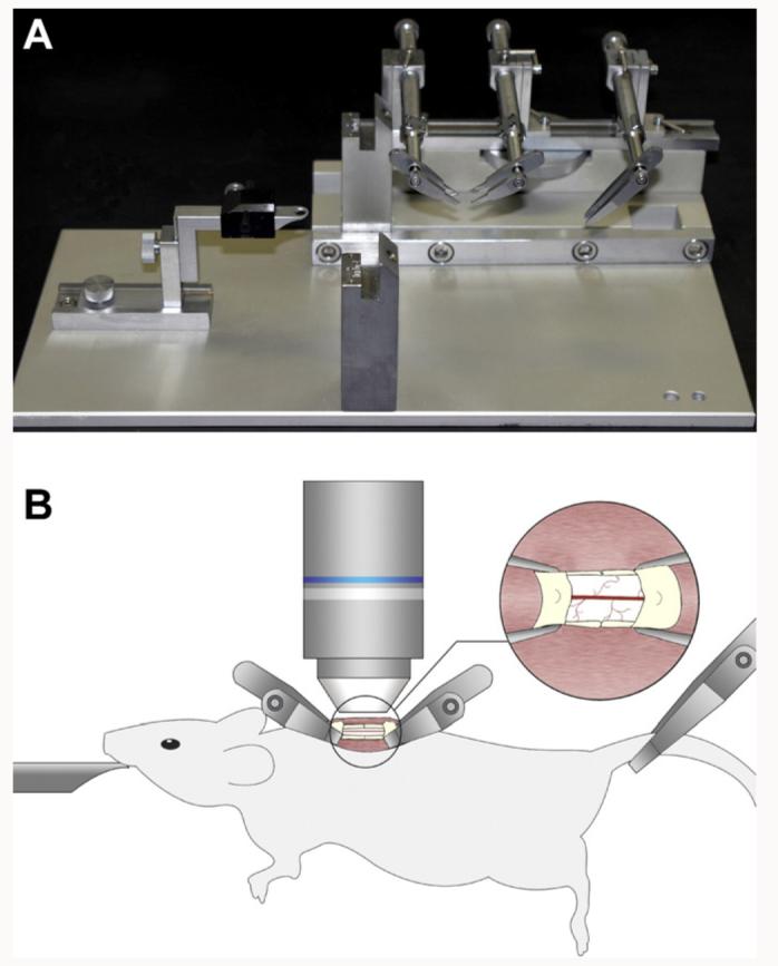
In vivo two-photon imaging of the stabilized mouse spinal cord. (A) A spinal stabilization device that could fit on a lowered microscope stage was built as shown here, using a steel base plate to support and align the STS-A Narishige compact spinal cord clamps and the MA-6N Narishige head holding adaptor. (B) Adult transgenic mice anesthetized with a KXA mix were positioned on the spinal stabilization device as shown here. A small retraction of the paravertebral muscles allowed the insertion of the fine tips of the clamping device and a laminectomy exposed the spinal cord. The entire device was placed in a temperature controlled chamber under the two-photon microscope and a heated water immersion lens dipped in a drop of ACSF was used for in vivo imaging of the fluorescently labeled spinal cord.