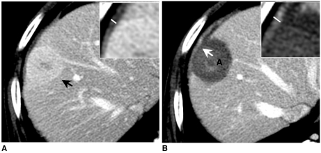
Fig. 2.
76-year-old man with hepatocellular carcinoma showing diaphragm swelling and enhancement.
A. Portal phase CT scan obtained prior to radiofrequency ablation shows enhancing nodule (black arrow) in segment VIII. White line = diameter of diaphragm thickness.
B. Portal phase CT scan obtained immediately after radiofrequency ablation shows ablation zone (A) with minimal amount of fluid collection. Swelling and enhancement of diaphragm is noted as thermal injury for abutting ablation zone (white arrow). White line = diameter of diaphragm thickness.

