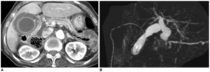
Fig. 14.
Choledochal cyst with anomalous union of pancreaticobile duct (Komi IA, Todani IC) combined cholecystitis.
A, B. Initial CT (A) image shows diffuse wall thickening with distension of gallbladder, suggesting acute cholecystitis. MR cholangiopancreatography (B) image shows fusiform dilatation of common bile duct, joined with pancreatic duct at right angle.

