Fig. 5.
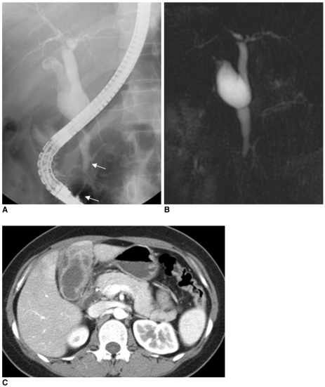
Choledochal cyst with anomalous union of pancreaticobile duct (Komi IIA, Todani IB) combined small cell cancer of gallbladder.
A-C. Endoscopic retrograde cholangiopancreatography (A) and MR cholangiopancreatography (B) images show fusiform dilatation of common bile duct, joined with pancreatic duct at acute angle, along with long common channel (arrows, 24 mm) and low lying cystic duct insertion with dilatation in proximal region. Irregular filling defect is noted in contrast filled gallbladder on endoscopic retrograde cholangiopancreatography image. CT image (C) shows irregular enhancing wall thickening of gallbladder. Pathologic diagnosis was confirmed to be small cell carcinoma.

