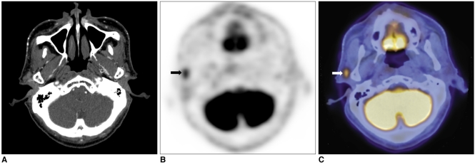
Fig. 1.
65-year-old male patient with sebaceous carcinoma in upper eyelid.
A. On contrast-enhanced CT, no definite lesion is detected around parotid gland.
B, C. On PET/CT, malignant lesion with high glucose uptake of SUV 4.7 is diagnosed at same site (arrows). According to fused image, malignant lesion is located around parotid gland.

