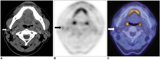
Fig. 2.
56-year-old male patient with sebaceous carcinoma in upper eyelid.
A. On contrast-enhanced CT, upper jugular lymph node behind angle of mandible is considered benign, with pattern of low enhancement and oval shape (arrow).
B, C. On PET/CT, same lesion showed asymmetrical glucose uptake with SUV 2.2 and is diagnosed as metastatic lymph node (arrows). As result, surgical fields were extended to include specific lymph node, which was pathologically proven to have metastatic lesion.

