
Figure 1.
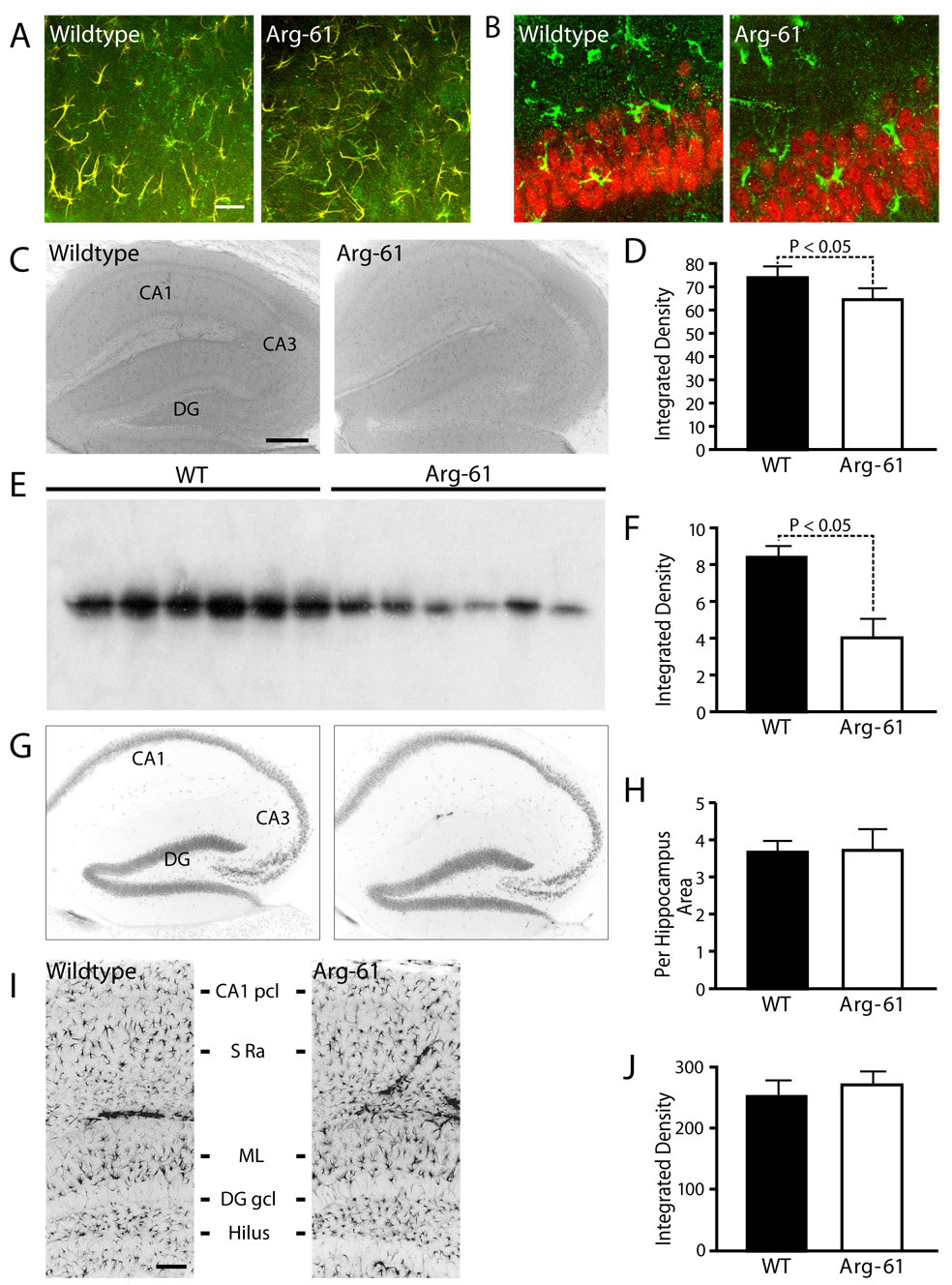
(A) Double immunostaining for apoE (green) and GFAP (red) showed co-localization (yellow) of apoE in astrocytes somas and larger cellular processes in hippocampus. (B) Double immunostaining for apoE (green) and NeuN (red) showed no co-localization. (C–F) ApoE expression and protein levels in the brains of 12-month-old WT and Arg-61 apoE mice. ApoE immunoreactivities in the hippocampus were assessed immunohistochemically (C), quantitated as immunochemical integrated density (D, n = 9 per group) and by western blot of mouse brain homogenates (E, F, n = 6 mice per group). (G) Distribution of NeuN-positive hippocampal neurons in the brain of WT and Arg-61 apoE mice, and (H) the relative numbers of neurons quantitated in selected CA1 areas of hippocampus (n = 9 mice for each group). (I) Distribution of GFAP-positive astrocytes in different layers of hippocampus (CA1 pcl – CA1 pyramidal cell layer, S ra – strata radiatum, ML – molecular layer, DG gcl – dentate gyrus granule cell layer) in the brain of WT and Arg-61 apoE mice, and (J) the GFAP immuno-reactivities were quantitated in the hippocampus (n = 10 for each group). Scale Bars: (A) 10 µm, (C) 250 µm, (I) 50 µm.