
Figure 2.
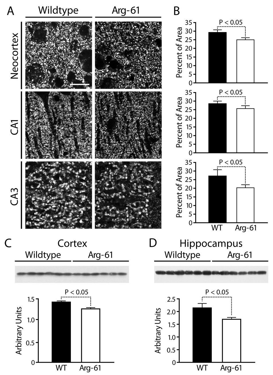
Synaptophysin expression in the brains of WT and Arg-61 apoE mice. (A, B) Immunostaining for synaptophysin and quantitation in the neocortex and the CA1 and CA3 areas of the hippocampus (n = 9 mice per group). (C, D) The protein levels of synaptophysin in the cortex and hippocampus were quantitated by western blot analysis (n = 6 mice per group). Scale Bar: 5 µm.