
Figure 4.
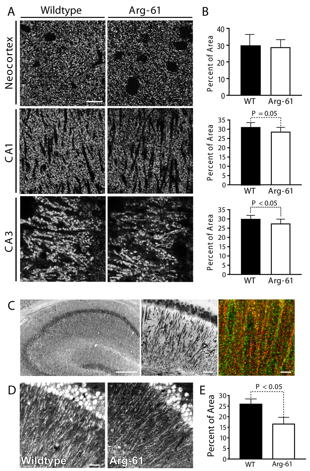
Presynaptic integrity accessed by Bassoon levels and determination of NLG1 positive dendrites and postsynaptic terminals in the brains of WT and Arg-61 apoE mice. (A) Immunostaining for Bassoon in neocortex and hippocampus and (B) quantitated by percent of immuno-reactive area (n = 9 per group). (C) Sample picture of immunostaining of hippocampal CA1 pyramidal neurons (left panel) and dendrites (central panel) by NLG1 in WT mouse brain. (C, right panel) Sample picture of WT mouse hippocampal CA1 region, double labeling postsynaptic terminals with GluR2/3 (green) and NLG1 (red), colocalization of GluR2/3 with NLG1 are shown in yellow and orange. (D) Immunostaining of positive postsynaptic terminals by NLG1 and (E) quantitation by percent of area of NLG1 immunoreactivity (n = 6 per group). Scale Bars: (A) 5 µm, (C) left panel 250 µm, central 50µm, right 5 µm, (D) 25 µm.