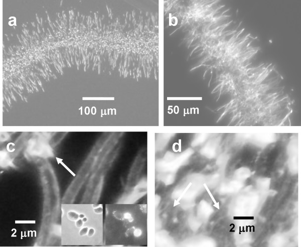
Figure 4.
Detached biofilm structure (3 h biofilms). All images were acquired using epi-fluorescence microscopy. a) Spurr's embedded section; the fluorescence originates from the preparation technique; b) Cryosection stained with calcofluor white; c, d) Cryosections stained for (1,3) β glucan; c) Region at the edge of the biofilm; arrow indicates extracellular material that is stained; inset is the planktonic positive control (left: transmitted image right: epi-fluorescence image); d) region in the interior of the biofilm; arrows indicate stained material that appears as strands.

