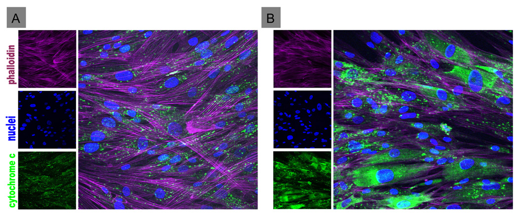
Figure 5. Effect of TRH on apoptosis of hIPCs as monitored by release of mitochondrial cytochrome C into the cytoplasm.
Uninfected hIPCs (A) or hIPCs infected with 3,000 particles of AdCMVmTRHR per cell (B) were incubated in serum-free medium without (A) or with 1 µM TRH (B) for 24 h and then fixed and stained for f-actin, cytochrome C and nuclei. TRHR-expressing cells not treated with TRH (not shown) were similar to uninfected hIPCs (A) demonstrating punctate cytochrome C staining indicating mitochondrial localization. TRHR-expressing hIPCs treated with TRH (B) showed dispersed cytoplasmic staining indicating leakage of cytochrome C from mitochondria.

