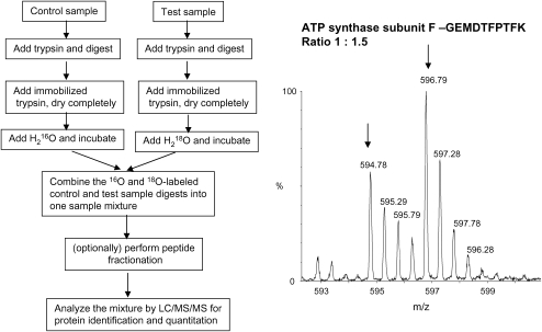
Figure 5.
Overview of 16O/18O labeling methodology. The left schematic diagram shows the flow of 16O/18O methodology, and the spectra on the right shows an example mass spectrum. The control was labeled with 16O and the test sample was labeled with18O with a ratio of 1:1.5, and this peptide GEMDTFPTFK is from ATP synthase subunit F. The MS spectrum is used courtesy of Shijun Sheng, Johns Hopkins University.

